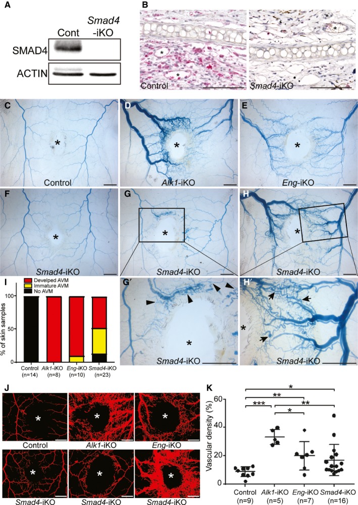
Figure 5.

Smad4 deficiency causes skin arteriovenous malformations (AVMs) in response to wounding in adults. A and B, Western blot (A) of wounded skin and immunostaining of wounded ears with anti‐SMAD4 antibodies demonstrate that the SMAD4 protein is diminished in Smad4‐inducible knockout (iKO) mice. ACTIN was used as a loading control. Scale bars in B=100 μm. C through H’, Latex dye–perfused vasculature surrounding the wound in the dorsal skin of control (C), Alk1‐iKO (D), Eng‐iKO (E), and Smad4‐iKO (F through H’) mice 6 days after tamoxifen treatment. While most of the Alk1‐iKO and Eng‐iKO mice displayed well‐developed AVMs, Smad4‐iKO mice exhibited a range of severity of skin AVMs: no AVM (F), immature AVMs (G and G’), and developed AVMs (H and H’). Arrows indicate fully developed AVMs and arrowhead marks show immature AVMs. The wound sites are indicated by asterisks. The scale bars indicate 2 mm. I, Percentage of categorized wound‐induced AVMs in hereditary hemorrhagic telangiectasia model animals. J, Blood vessels containing the latex dye are processed for quantification. Representative converted images to quantify the vascular area containing latex. The wound sites are indicated by asterisks. The scale bars indicate 2 mm. K, Quantification of vascular density per a given area. All data represent mean±SD. *P<0.05, **P<0.01, and ***P<0.001. Cont indicates control.
