
Figure 1.
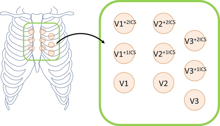
Position of 9 right precordial ECG leads analyzed in this study. In this study, V1 to V3 leads positioned in the standard and upper 1 and 2 intercostal spaces (+1ICS and +2ICS, respectively) were recorded for evaluation of the number and amplitude of type 1 ECG.