
Figure 3.
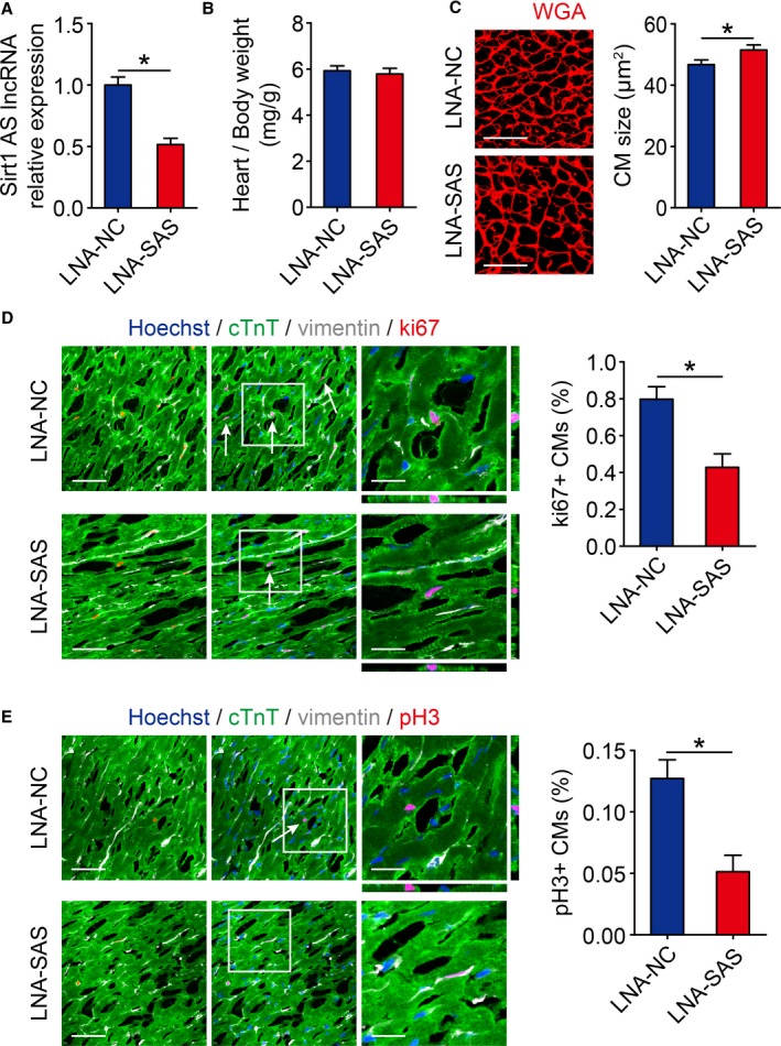
Inhibition of Sirt1 antisense (AS) long noncoding RNA (lncRNA) suppresses cardiomyocyte (CM) proliferation in vivo. A, Real‐time quantitative PCR analyses of Sirt1 AS lncRNA levels in locked nucleic acid–control (LNA‐NC) or LNA–Sirt1 AS lncRNA (SAS) injected neonatal mouse hearts 10 days after injection (n=6 mice per group). B, Ratios of heart weight/body weight in neonatal mouse hearts injected with LNA‐NC or LNA‐SAS (n=5 mice per group). C, Wheat germ agglutinin (WGA) staining of heart sagittal sections for LNA‐NC and LNA‐SAS adult mice hearts and quantification of CM size (167 CMs from 5 mice in LNA‐NC group, 167 CMs from 5 mice in LNA‐SAS group). Bar=25 μm. D, Immunofluorescence of ki67 in neonatal hearts injected with LNA‐NC or LNA‐SAS and quantification of ki67‐positive CMs (1627 CMs from 5 mice in LNA‐NC group, 1671 CMs from 5 mice in LNA‐SAS group). Ki67‐positive CMs were indicated by arrows. Bars: 50 μm (left panels); 20 μm (right panels). E, Immunofluorescence of pH3 in neonatal hearts injected with LNA‐NC or LNA‐SAS and quantification of pH3‐positive CMs (6244 CMs from 5 mice in LNA‐NC group, 7416 CMs from 5 mice in LNA‐SAS group). pH3‐positive CMs were indicated by arrows. Bars: 50 μm (left panels); 20 μm (right panels). Statistical significance was calculated using 2‐tailed unpaired Student t test in A through E. Data represent mean±SEM. *P<0.05.