
Figure 5.
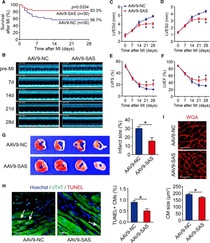
Sirt1 antisense (AS) long noncoding RNA (lncRNA) induces cardiac regeneration after myocardial infarction (MI) in adult mice. A, Kaplan‐Meier survival curves in adeno‐associated virus 9–negative control (AAV9‐NC) and AAV9–Sirt1 AS lncRNA (SAS) adult mice after MI (n=30 mice per group). B through F, The M‐mode ultrasonic cardiography change of mice hearts detected by echocardiography in AAV9‐NC and AAV9‐SAS adult mice at pre‐MI and 7, 14, 21, and 28 days after MI and quantitative analyses of left ventricular internal diameter at end diastole (LVEDd; C), left ventricular internal diameter at end systole (LVESd; D), left ventricular fractional shortening (LVFS; E), and left ventricular ejection fraction (LVEF; F) (n=5 mice per group). G, Triphenyl tetrazolium (TTC) staining of mice ventricular cross sections in AAV9‐NC and AAV9‐SAS adult mice 28 days after MI and quantitative analyses of TTC staining infarct size (n=5 mice per group). H, Cell apoptosis was detected by terminal deoxynucleotidyl transferase–mediated dUTP nick end labeling (TUNEL) staining in AAV9‐NC and AAV9‐SAS adult mice 28 days after MI and quantitative analyses of TUNEL‐positive cardiomyocytes (CMs; 1343 CMs from 5 mice in AAV9‐NC group, 1387 CMs from 5 mice in AAV9‐SAS group). TUNEL‐positive CMs were indicated by arrows. Bar=20 μm. I, Wheat germ agglutinin (WGA) staining in AAV9‐NC and AAV9‐SAS adult mice 28 days after MI and quantitative analyses of CM size (139 CMs from 5 mice in AAV9‐NC group, 139 CMs from 5 mice in AAV9‐SAS group). Bar=25 μm. Statistical significance was calculated using log‐rank (Mantel‐Cox) test in A, 1‐way ANOVA followed by least significant difference post hoc test in C through F, and 2‐tailed unpaired Student t test in G through I. Data represent mean±SEM. *P<0.05.