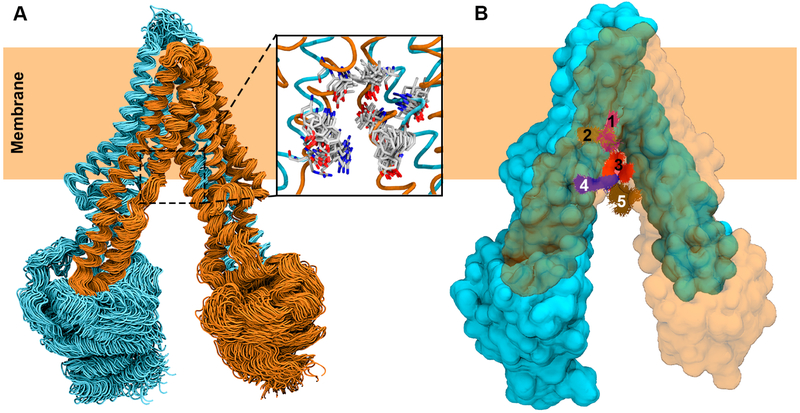
Figure 5.
Ensemble docking of a drug substrate into the translocation lumen of P-gp. (A) MD simulation-generated ensemble of conformations used in molecular docking. Two pseudosymmetric halves of P-gp are colored in cyan and orange, respectively. Only 100 out of 1000 conformations used in ensemble docking are shown. A set of translocation path residues is shown to display the diverse side chain conformations represented by the MD-generated ensemble (inset). (B) Top 5 RMSD-based clusters of docked poses of the drug molecule are shown in different colors with P-gp in surface representation. One half of the protein is shown transparent to clearly show the clusters of the docked drug molecule.

