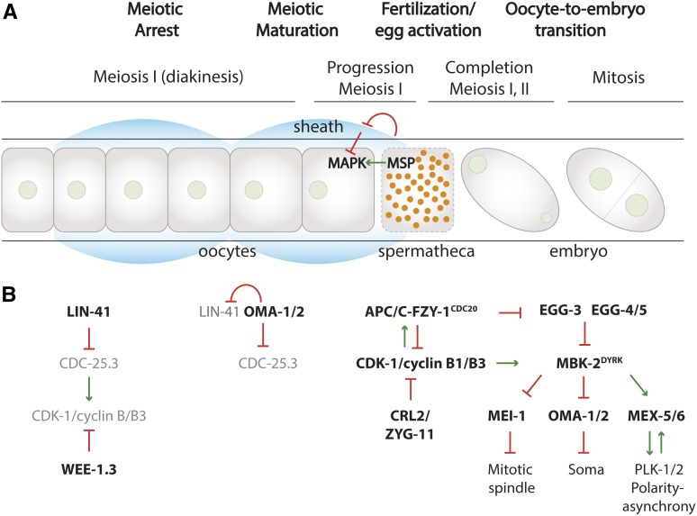
Figure 3.
Meiotic arrest, oocyte maturation, fertilization, and the oocyte-to-embryo transition in the hermaphrodite germline. (A) Schematic illustration of the successive stages from meiosis I-arrested oocytes in the gonad arm (left) to mitotic embryos in the uterus (right). Nuclei are green, sperm are orange. (B) Diagram of various regulators that mediate the cell cycle transitions in time, from left to right. Initially, WEE-1.3 and LIN-41 are thought to antagonize CDK-1 activation. See text for further information.

