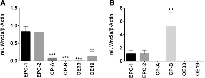
Fig. 1.
Expression of the extracellular ligands Wnt3a and Wnt5a along Barrett’s sequence. Expression of Wnt3a (a) and Wnt5a (b) was analyzed in EPC-1 and EPC-2 (squamous esophageal epithelium), CP-A (metaplastic epithelium), CP-B (epithelium with high-grade dysplasia), OE33 and OE19 (esophageal adenocarcinoma) cells by quantitative Real time RT-PCR. Highest Wnt3a expression was detected in EPC-1 and EPC-2 (a). Wnt5a was also expressed in EPC-1 and EPC-2, but highest levels of Wnt5a were found in CP-B (b). OE33 and OE19 expressed only marginal levels of Wnt3a and Wnt5a (a, b). Normalization was done with β-Actin. Values are shown as mean ± S.E.M. (One-way-ANOVA with Bonferroni correction, * - p < 0.05, ** - p < 0.01, *** - p < 0.001 compared to EPC-1)

