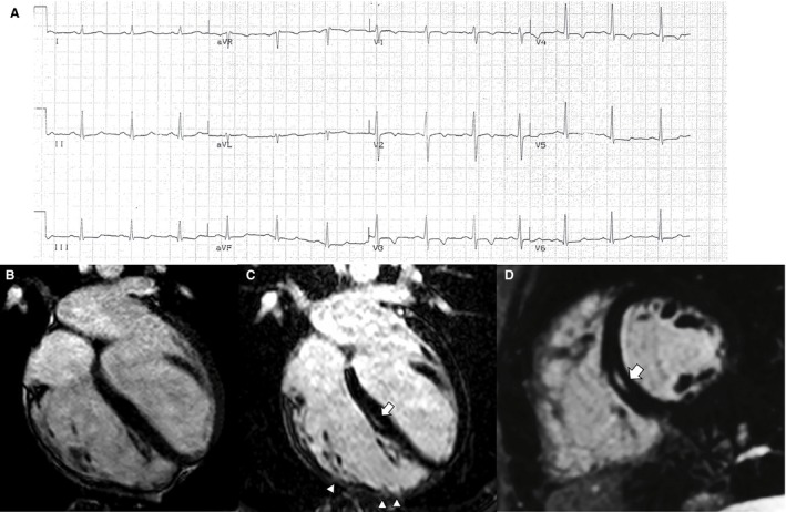
Figure 1.

Electrocardiographic and contrast‐enhanced cardiac magnetic resonance (CMR) findings of a representative case of right‐dominant (classic) arrhythmogenic cardiomyopathy variant. A, Basal ECG showing T‐wave inversion in right precordial leads (V1‐V4). B, End‐diastolic frame of cine CMR sequence in long‐axis 4‐chamber view showing a dilated right ventricle (end diastolic volume, 127 mL/m2) with a severely reduced ejection fraction (25%). The postcontrast orthogonal images in long‐axis (C) and short‐axis (D) views show late gadolinium enhancement as midwall stria in the midseptum (white arrow). In C, late gadolinium enhancement is also visible in the anterolateral, mid, and apical regions of the right ventricular wall, with segmental transmural involvement (white arrowheads) associated with regional dyskinesia (not shown).
