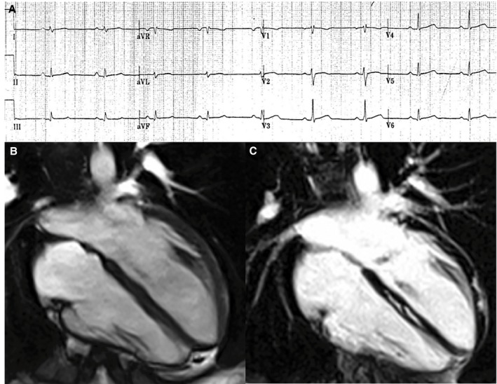
Figure 3.

Electrocardiographic and contrast‐enhanced cardiac magnetic resonance (CMR) findings of a representative case of left‐dominant arrhythmogenic cardiomyopathy variant in a patient with a desmoplakin‐gene mutation and a history of sustained ventricular tachycardia. A, Basal ECG showing low QRS voltages (<0.5 mV) in limb leads. B, End‐diastolic frame of cine CMR sequence in long‐axis 4‐chamber view showing normal cavity size and function of both ventricles. C, Postcontrast image showing myocardial fibrosis in the form of stria of late gadolinium enhancement in the epicardium of the left ventricular lateral wall (arrowheads) and midmural layer of the interventricular septum (arrows).
