
Figure 3.
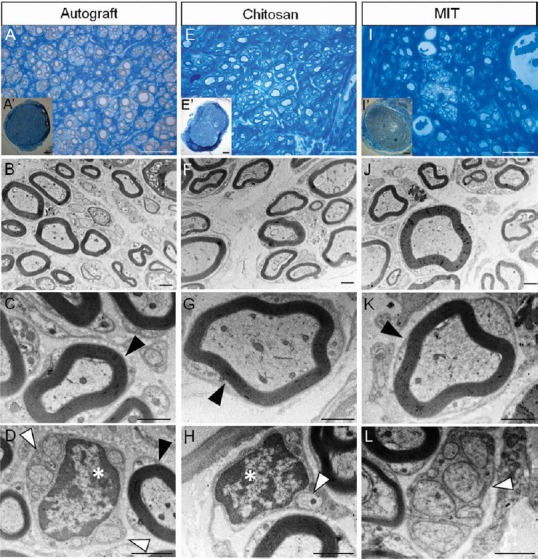
Morphology of regenerated nerve fibers 4 months after median nerve repair.
Representative high and low magnification light microscopy images of toluidine blue-stained semi-thin cross sections of nerve repaired with autograft (A and A’), nerve repaired with hollow chitosan conduit (E and E’) and nerve repaired with MIT (I and I’). Scale bars: 20 µm in A, E, I; 100 µm in A’, E’, I’. Representative high magnification transmission electron microscopy images of ultrathin cross sections of nerve repaired with autograft (B–D), with hollow chitosan conduit (F–H) and with MIT (J–L). Scale bars: 1 µm in B–D, F–H, and J–L. Black arrowheads: myelinated fibers with well-defined axoplasm and well-organized myelin sheaths; white arrowheads: unmyelinated fibers; white asterisks: Schwann cell nuclei. All experimental groups display regenerated fibers with many regrowing myelinated and unmyelinated fibers. MIT: Muscle-in-tube.