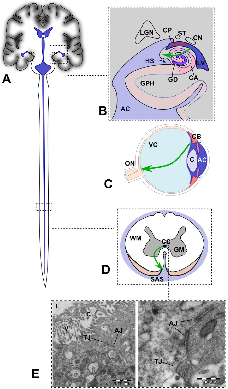
Figure 2.

Sites of focal rupture of the fluid-brain barrier (FBB) in the central nervous system (not at scale).
(A) Schematic representation of the ventricular system including the central canal containing CSF (in blue). (B) Drawing of a coronal section of the brain though the hippocampus. The green arrow indicates anomalous flow of CSF through a focal break of the FBB, as postulated in AD. Adapted from Duvernoy et al. (2013). (C) Drawing of a midsagital section of the eye globe showing the anterior and posterior chambers filled with aqueous humor (in blue). The green arrow indicates anomalous posterior flow of aqueous, as postulated in glaucoma. (D) Drawing of an axial section of the spinal cord showing the central canal and the subarachnoid space filled with cerebrospinal fluid. The green arrow indicates anomalous flow of CSF through a focal break of the FBB, as postulated in amyotrophic lateral sclerosis. (E) Electron microscopy micrography showing TJ and AJ in the ependymal cells lining the CC of the spinal cord of the rat. In humans, the presence of TJs in the CC is transient during development and irregularly distributed in adults. AC: Ambient cistern; CA: cornu ammonis; CN: caudate nucleus (tail); CP: choroidal plexus; GD: girus dentatus; GPH: parahippocampal girus; HS: hippocampal sulcus; LGN: lateral geniculate nucleus; LV: lateral ventricle; ST: stria terminalis; C: cristaline lens; CB: ciliary body; ON: optic nerve; VC: vitreous body; CC: central canal; GM: grey matter; SAS: subarachnoid space; WM: white matter; TJ: tight junctions; AJ: adherent junctions.
