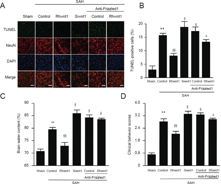
Figure 5.

Effect of rhwnt1, siwnt1, and anti-Frizzled1 treatment on cortical cell apoptosis and brain edema after experimental SAH.
(A) Immunofluorescence of TUNEL (green) and DAPI (blue) staining. Arrows indicate apoptotic cells in brain tissue. Scale bars: 32 μm. (B) Variation of apoptotic cell percentage in different groups. (C) Variation of brain water content percentage in different groups. Data are shown as the mean ± SEM (n = 6; one-way analysis of variance followed by Scheffé F post hoc test). **P < 0.01, vs. sham group; §P < 0.05, §§P < 0.01, vs. SAH + control group; ΔP < 0.05, vs. SAH anti-Frizzled1 control group. SAH: Subarachnoid hemorrhage; TUNEL: terminal deoxynucleotidyl transferase-mediated dUTP nick end labeling; DAPI: 4′,6-diamidino-2-phenylindole.
