Figure 6.
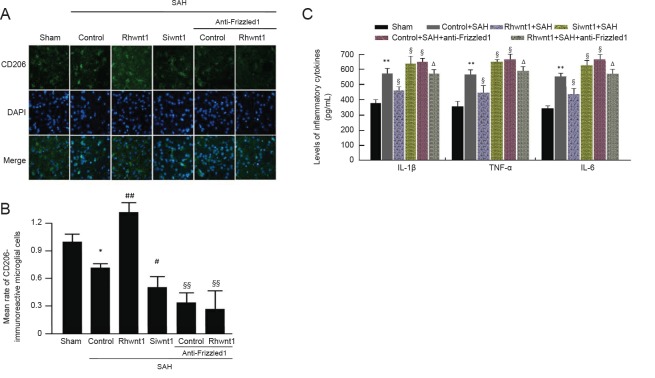
Effect of rhwnt1, siwnt1, and anti-Frizzled1 treatment on microglia conversion into M2-type and inflammatory cytokine release after experimental SAH.
(A) Double immunofluorescence analysis by fluorescence microscopy (original magnification, 100×) with CD206 antibody (green) and nuclei fluorescently stained with DAPI (blue). Arrows indicate CD206-immunoreactive cells. (B) Mean rate of CD206-immunoreactive microglial cells in the sham group was used as the standard. (C) Levels of inflammatory cytokines (IL-1β, IL-6, and TNF-α) measued by ELISA method. Data are shown as the mean ± SEM (n = 6; one-way analysis of variance followed by Scheffé F post hoc test). *P < 0.05, **P < 0.01, vs. sham group; §P < 0.05, vs. SAH + control group; ΔP < 0.05, vs. SAH + anti-Frizzled1 control group. SAH: Subarachnoid hemorrhage; DAPI: 4′,6-diamidino-2-phenylindole; IL: interleukin; TNF: tumor necrosis factor; ELISA: enzyme linked immunosorbent assay.

