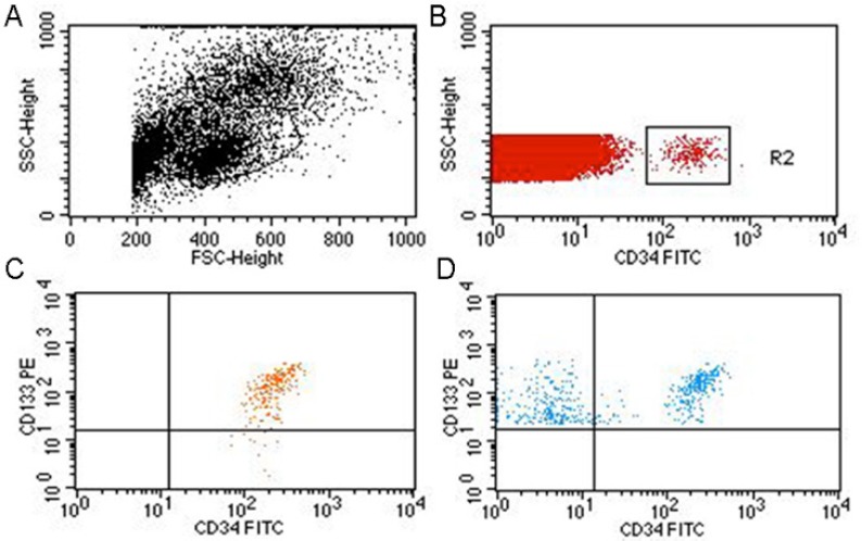
Figure 1.

Flow cytometry for detection of CD34- and CD133-positive cells.
Cells were first gated based on size and forward scatter. The black-and-white chart shows exclusion of platelets, dead cells, cell masses and cell fragments. The boxed area shows mononuclear cells to be analyzed (A). The gated cells were then collected via an FITC CD34-positive gate (boxed area in B). Mouse IgG1 conjugated with FITC or PE were used as isotype controls (the orange signal in C). The double-gated cells were then examined for CD133 and CD34 double-positive cells. The blue cells in the upper right quadrant are CD34 and CD133 double-positive cells (D). FITC: Fluorescein isothiocyanate; PE: phycoerythrin.
