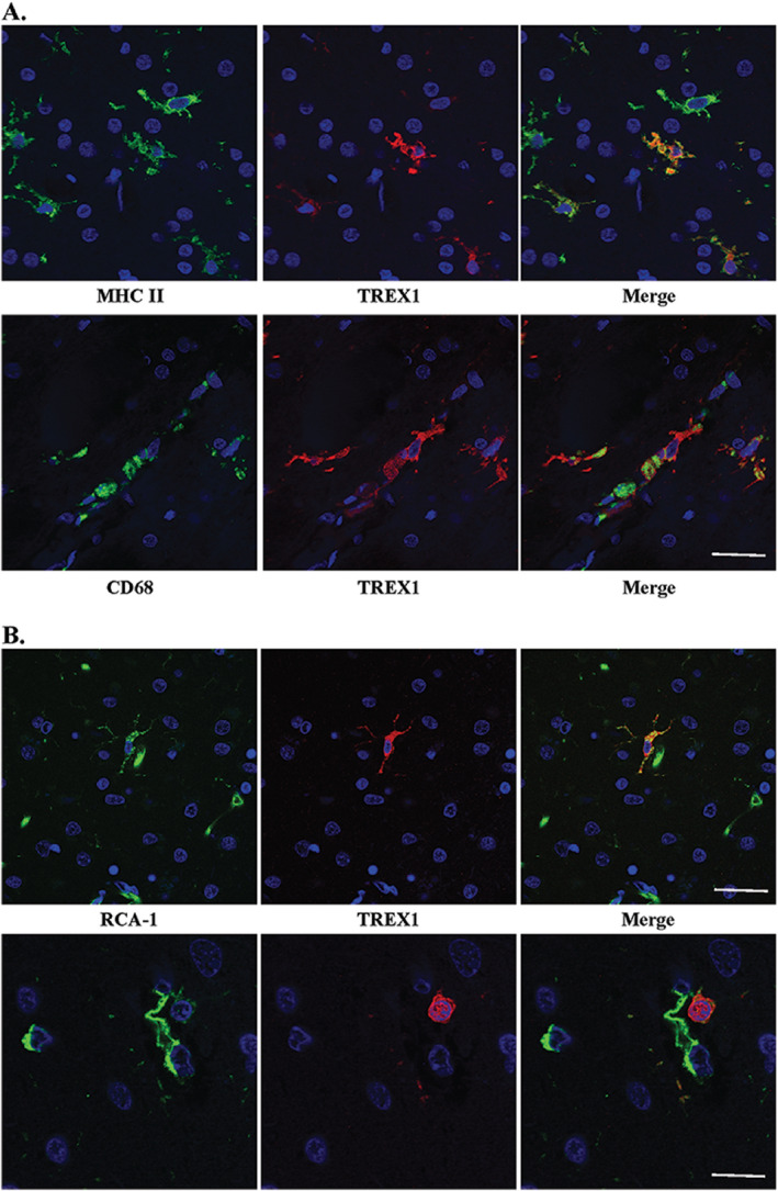
Figure 4.

TREX1 positive cells are microglia and/or macrophages. A. Dual staining of frozen tissue from a case of RVCL with anti‐MHC II (top panel, green) and anti‐CD68 (bottom panel, green), microglial markers, and anti‐TREX1 (red). Nuclei are counterstained with TO‐PRO‐3 (blue). Scale bar represents 28 μm. B. Two examples (upper and lower panels) of dual staining of formalin‐fixed, paraffin‐embedded human brain tissue from a normal control with RCA‐1 (left panel, green), a microglial/macrophage and endothelial cell marker, and anti‐TREX1 (red). Nuclei are counterstained with TO‐PRO‐3 (blue). Some cells stained with RCA‐1 but not TREX1 are endothelial cells based on their morphology. Scale bars represent 28 μm on top panel and 14 μm on bottom panel.
