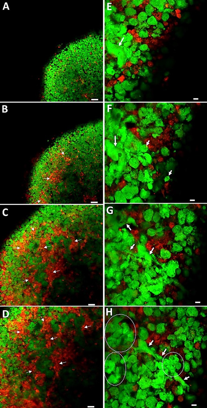
Figure 3.

Confocal microscopy images. A–D) Visualization of consecutive layers through a d 8 coculture cell pellet construct of HBMSCs (Vybrant, green) and HUVECs (Vybrant Dil, red). Snapshot images throughout the cell pellet constructs from the outside (A, E) to the inside (D, H). HBMSCs were observed predominantly in the outer layer of the pellet (A), with HUVECs increasing in number toward the center of the pellet (B–D). Arrows depict areas of HUVEC aggregation. E–H) Higher-magnification images depicting cell positions and migration within consecutive layers of the cell pellet construct. E, F) Visible variations in morphology of HBMSCs (arrows). G, H) Elongation of HBMSCs (arrows). H) Clusters of HBMSC aggregates (dotted lines). Scale bars, 50 µm (A–D); 10 µm (E–H).
