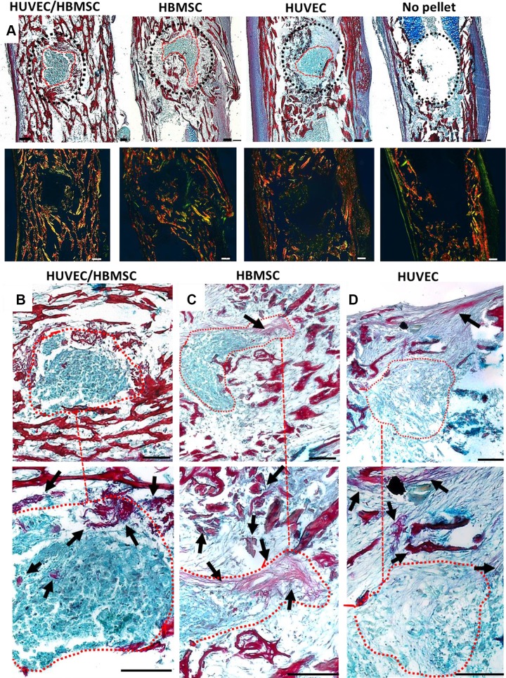
Figure 5.
Representative histology images depicting cell pellet construct integration (circled red) within femoral defects stained with Alcian Blue (proteoglycans)/Sirius Red (collagen). A) Overview of pellet integration in all treatments and the no-pellet control sample. A dotted black line surrounds the defect area. Images underneath depict birefringence imaging of collagen fiber distribution (thick collagen fibers shown in orange-red; thin collagen fibers shown in green). B–D) Magnified images of the pellet implant of each treatment group; higher-magnification images of the same area below (dotted line). Femur drill defects containing remnants of HUVEC/HBMSC coculture pellet (B), HBMSC pellet (C), and HUVEC pellet (D). Arrows show collagen formation within and around the defect area. Scale bar, 100 µm.

