Figure 1.
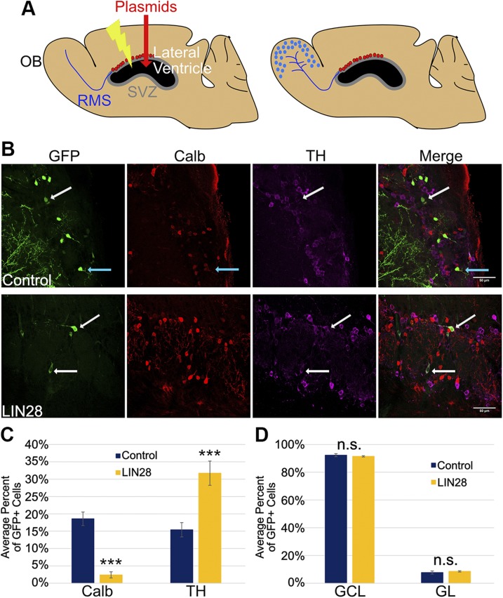
LIN28 alters cell fate in the GL of the OB at 21 DPE. A) Schematic of a sagittal mouse brain section depicting the electroporation protocol. A) At 0 DPE (left), plasmids are injected into the lateral ventricle, and then electrical impulses are administered so the DNA is incorporated in the NSCs of the SVZ (red dots). The NSCs of the SVZ produce progeny that migrate through the RMS to the OB at 14 DPE or later (right). B) Representative ×40 micrographs showing GFP (green), Calb (red), and TH (purple) immunostaining in the GL. Upper: control. Lower: LIN28. White arrows depict GFP/TH+ neurons; teal arrows depict a GFP/Calb+ neuron. Original scale bars, 50 μM. C) Bar graph depicting the average percent of GFP+ cells positive for Calb or TH at 21 DPE; n = 9 slices (control), n = 10 slices (LIN28::GFP). D) The average percent of GFP+ cells present in either the GCL or GL layers of the OB at 21 DPE; n = 7 slices. C, D) Data presented as means ± sem. ***P ≤ 0.005 vs. control, Student’s t test. N.s., not significant.

