Figure 3.
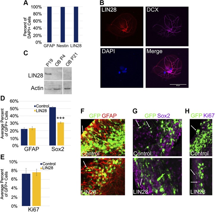
LIN28 is expressed in NPCs and at 3 DPE, affects the cell types of the SVZ differently. A) Percent of explanted primary DAPI+ NSCs positive for GFAP, Nestin, and LIN28. B) Micrographs (×40) depicting immunostaining of explanted, differentiated NSCs derived from the SVZ. LIN28 (red), DCX (purple), and DAPI (blue). Original scale bar, 50 μM. C) Western blot of LIN28 protein expression in undifferentiated P19 cells (positive control) and the OB in mice at postnatal d 4 and 21; n = 4 mice at each time point. Actin was used as a loading control. D) Bar graph showing the average percent of GFP+ cells positive for GFAP, a marker for NSCs and Sox2, a marker for NPCs at 3 DPE; n = 8 slices (control-GFAP), n = 7 slices (LIN28::GFP-GFAP), and n = 12 slices (Sox2). E) Bar graph depicting the percent of GFP+ cells positive for Ki67, a marker for active proliferation at 3 DPE; n = 12 slices (control), n = 9 slices (LIN28::GFP). F–H) Representative ×30 micrographs depicting immunostaining of NSCs (GFAP, red) (F), NPCs (Sox2, purple) (G), and actively proliferating cells (Ki67, purple) (H). Arrows depict examples of positive cells in each stain. A, D, E) Data presented as means ± sem. ***P ≤ 0.005 vs. control, Student’s t test.

