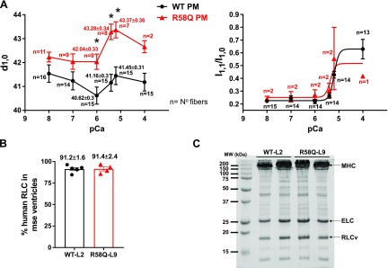
Figure 3.
A) IFS and I1,1/I1,0 as a function of pCa (8, 7, 6, 5.4, 5.2, and 4) in skinned PM fibers from left ventricles of 5–7-mo-old R58Q (2F, 4M) and WT (4F, 3M) mice. The sarcomere length (SL) was adjusted to 2.1 μm. Data are the average ± sem fibers, *P < 0.05 (Student’s t test). B). Mass spectrometry data (average ± sd) showing expression of the human RLC in the ventricles of WT (n = 6 samples) and R58Q (n = 4 samples) mice. C) Representative SDS-PAGE image of myosin purified from heart ventricles of WT and R58Q mice.

