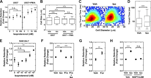
Figure 1.
β-AR signaling activation reduces the deformability of macrophages. A) Retention of monocyte (U937) and macrophage (U937+PMA) cells was measured using parallel microfiltration after isoproterenol treatment for 24 h. Retention is measured by the mass of cell suspension that remains in the top well after applying 2 kPa pressure for 30 s relative to the initial mass. Higher retention indicates that cells are more likely to occlude the 5 µm pores. B) Cell size distribution of monocytes and macrophages in vehicle control (Veh) and isoproterenol (Iso)-treated groups. C) Density scatterplot shows data from quantitative deformability cytometry. We measure cell size and transit time, or the timescale for macrophages to pass through the 5 × 10 μm gap of a microfluidic channel (n > 1600). D) Boxplot shows the lower and upper quartiles with median (line). Whiskers show the 10–90th percentiles (P < 0.0001). E) Isoproterenol effects on mouse macrophage (RAW 264.7) cells. F–H) Relative retention measured by PMF following 24 h treatment with Iso (100 nM) and the β-blocker propranolol (Pro) (10 μM) (F). Pro+Iso indicates cotreatment. Adenylyl cyclase activator forskolin (For; 10 μM) (G). Calcium chelator BAPTA-AM [1,2-Bis(2-aminophenoxy)ethane-N,N,N′,N′-tetraacetic acid tetrakis-acetoxymethyl ester] (10 μM) (H). One-way ANOVA test with Tukey’s multiple comparison test. *P < 0.05, **P < 0.01, ***P < 0.001; n.s., not significant. Results represent data from at least 3 independent experiments (N = 3).

