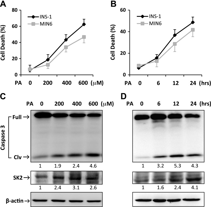
Figure 1.
PA induced apoptotic cell death and SK2 expression in β-cells. MIN6 and INS-1 cells were treated for 24 h with PA at increasing concentrations as indicated (A, C) or with PA at 400 μM for the indicated time points (B, D). A, B) The treated cells were costained with propidium iodide (PI) and annexin V–FITC (AV) followed by flow cytometric analysis. “Cell death” refers to the percentage of cells encompassing both AV single-positive and AV/PI double-positive cells. Error bars represent sd of the mean value from 3 independent experiments. C, D) Levels of full-length/cleaved (Clv) caspase-3 and SK2 expression were determined by Western blotting. Numbers below lanes indicate band intensity relative to that of the loading control β-actin. The blots represent 4 independent experiments.

