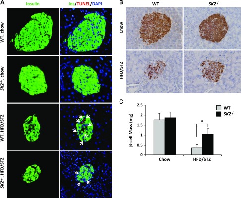
Figure 7.
SK2 deficiency protected β-cells against apoptosis in HFD/STZ-treated mice. A) Representative images show the pancreatic sections from control or HFD/STZ-treated WT and SK2-deficient mice, fluorescence stained for insulin (green), TUNEL (red), and DAPI (blue). Arrowheads indicate apoptotic (TUNEL-positive) β-cells. B) Representative images show sections immunohistochemistry stained for insulin. C) β-Cell mass was determined based on insulin staining according to pancreas weight (milligrams) × islets per field area × β-cell numbers per islet area. Photomicrographs were taken of individual fields. Original magnification, ×40. At least 5 fields were taken from each section, and at least 3 pancreatic sections were counted from each animal. Data are expressed as mean ± sem. *P < 0.001.

