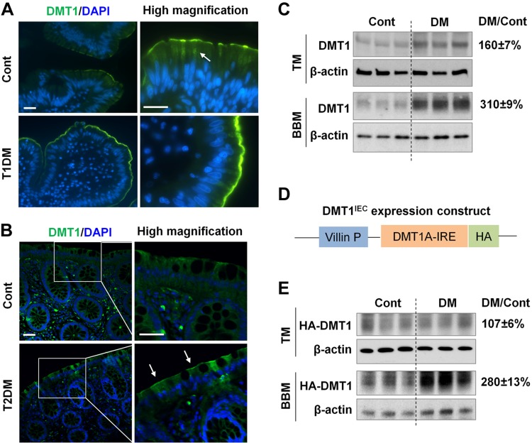
Figure 3.
Microvillus membrane expression of DMT1 is increased in diabetic humans and mice. A) Human jejunal biopsies were obtained from patients with type 1 diabetes (T1DM) and from healthy controls (Cont). DMT1 expression (green) was assessed by immunofluorescence staining with anti-human DMT1 antibody. Nuclei (blue) were stained with DAPI. Arrow, cytoplasmic localization of DMT1. Scale bar, 20 μm. B) Representative images showing DMT1 expression in human colonic biopsies from patients with T2DM and from Cont. Arrow, luminal membrane expression. Scale bar, 20 μm. C) DMT1-IRE expression in the duodenal total membrane (TM) and BBM of Cont and diabetic WT mice was determined by Western blotting with anti-rat DMT1 antibody. D) Transgenic DMT1IEC mice express HA-tagged human DMT1A-IRE isoform specifically in IECs using villin gene promoter. E) The expression of HA-DMT1A-IRE (HA-DMT1) in the duodenal TM and BBM of Cont and diabetic (DM) DMT1IEC mice was determined by immunoblotting with anti-HA antibody. β-actin was used as a loading control. The relative fold changes (DM/Cont) of DMT1 or HA-DMT1 expression in DM vs. Cont mice are shown on the right. Data are expressed as means ± se (n = 6).

