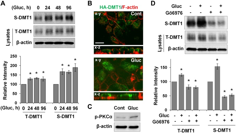
Figure 5.
High glucose induces PKCα-dependent membrane accumulation of DMT1 in cultured intestinal epithelial cells. A) Filter-grown T84 cells were treated with 20 mM d-glucose, and plasma membrane DMT1 expression was determined by cell-surface biotinylation assay. Surface (S) and total (T) DMT1 expression was determined by Western blotting with anti-human DMT1 antibody. Relative changes in DMT1 expression are expressed as means ± se (n = 3 independent experiments). B) Representative confocal images of the x–y and x–z planes show localization of HA-DMT1 (green) in glucose-treated (Gluc) and untreated (Cont) T84/HA-DMT1 cells. F-actin (red) was stained with phalloidin to outline the plasma membranes. Scale bar, 10 μm. C) p-PKCα expression in Gluc-treated and Cont T84 cells was determined by immunoblotting. D) T84 cells were pretreated for 30 min with Gö 6976 (1 μM) before glucose treatment for 24 h. T- and S-DMT1 expression was evaluated by immunoblotting, and the relative changes in their expression are shown as means ± se (n = 3). *P < 0.01 compared with the untreated control (set at 100).

