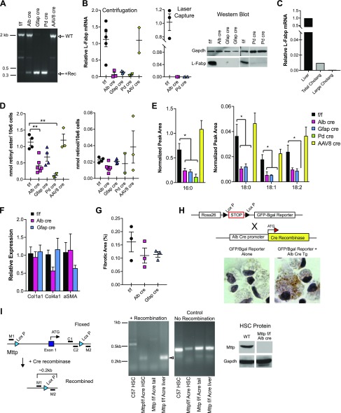
Figure 2.
HSC-specific deletion of L-Fabp. A) PCR amplification (P1/P2 primers) of genomic DNA from stellate cells from L-Fabp f/f mice, Alb Cre, Gfap Cre, and Pd Cre mice and from L-Fabp f/f mice injected with AAV8 Tbg cre. Note the partial deletion of the Lox P–flanked region in HSCs from mice expressing Alb Cre but not AAV8 Tbg Cre. Based on differences in PCR product size and staining intensity, recombination efficiency is estimated to be ∼50%. B) L-Fabp mRNA (left, middle) and protein (right) expression in HSCs isolated by differential centrifugation (left, right) or laser capture microdissection (middle) from L-Fabp f/f and Alb Cre, Gfap Cre, and Pd Cre mice. For Western blot (right), expression of Gapdh is shown as a loading control. C) Relative expression of L-Fabp mRNA in pools of isolated C57BL/6N cholangiocytes (total or large) expressed relative to L-Fabp mRNA levels in whole liver (C57BL/6J mice). D) Total retinyl ester (left) and retinol levels in freshly isolated HSC pools from L-Fabp f/f, Alb Cre, Gfap Cre, Pd Cre and AAV8-Tbg Cre injected L-Fabp f/f mice (n = 2–6 pools per genotype). E) Relative abundance of individual retinyl ester species. *P < 0.05, **P < 0.01, ***P < 0.001. F) Baseline fibrogenic gene expression in liver of chow-fed female mice (∼12 wk, n = 4–7/genotype). G) Sirius red–stained area in liver tissue from chow-fed female mice (n = 3/genotype). H) To examine whether Albumin Cre promotes recombinase expression in HSC, Alb cre Tg mice were crossed with mice expressing a floxed GFP/β-gal Cre reporter transgene (top). GFP/β-gal protein is expressed after deletion of intervening LoxP flanked stop sequences by Cre. Expression of GFP was detected by immunostaining in HSCs isolated from Alb Cre Tg mice (right) but not in HSCs expressing only the reporter. I) Alb-Cre induces partial recombination of Mttp gene. Schematic diagram (left) shows the location of primers spanning the deleted region (M1/M2, to detect recombination) and within the deleted region (C1/C2, control). Partial recombination of Mttp locus is detected in HSCs isolated from Mttp f/f Alb Cre mice, with reduced amplification of the control PCR product (middle panel) and no expression of Mttp protein in HSCs (right). Gapdh is shown as a loading control.

