Figure 3.

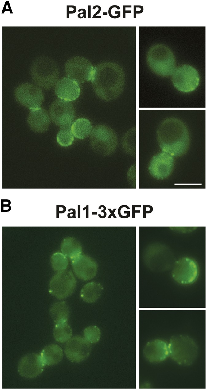
Pal2 and Pal1 localize to to cortical patches. (A) Pal2-GFP (SL7455); (B) Pal1-3xGFP (SL7335) were imaged by fluorescence microscopy. Shown are images from a single medial plane of a z-stack. Scale bar: 5μm.

Pal2 and Pal1 localize to to cortical patches. (A) Pal2-GFP (SL7455); (B) Pal1-3xGFP (SL7335) were imaged by fluorescence microscopy. Shown are images from a single medial plane of a z-stack. Scale bar: 5μm.