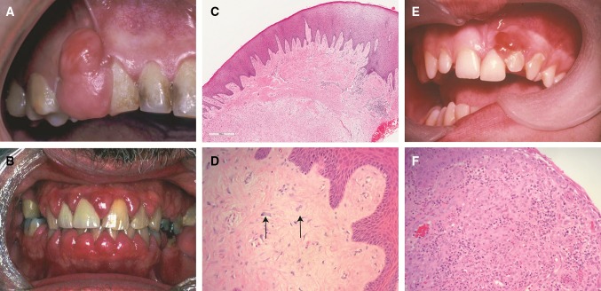
Fig. 1.
a A large fibrous epulis on maxillary gingiva. b Widespread fibrous gingival enlargement on a patient on cyclosporine therapy. c Histological image of a nodule of fibrous hyperplasia of the gingiva (H&E, Overall magnification × 20). In this case, the collagen varies from superficially hyalinised to more edematous in deeper tissues. d Histological image showing large stellate fibroblasts in a giant cell fibroma (H&E, overall magnification × 200). e An ulcerated vascular lesion on the maxillary gingiva of a pregnant patient in mid-trimester. f The histology of a vascular epulis/pyogenic granuloma shows attenuated or ulcerated epithelium with an underlying endothelial proliferation. This may have a lobular pattern (H&E, overall magnification × 200)

