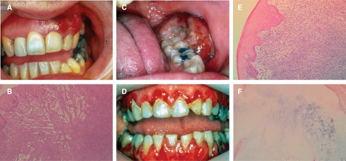
Fig. 4.
a Red, nodular swelling affecting the facial gingiva above the left maxillary canine and lateral incisor in Kaposi Sarcoma. b Streams of spindled cells with slit-like vessels and lymphangiomatous pattern superficially in Kaposi’s sarcoma (H&E, overall magnification × 4). c Non-Hodgkin lymphoma presenting as an ulcerated swelling affecting the posterior left retromolar region. d Generalized erythema and swelling affecting the gingiva in a case of AML. e Connective tissue effaced by sheets of atypical myeloid cells in AML (H&E, overall magnification × 20). f Chondrosarcoma classically has a lobular architecture, with blue-grey cartilaginous matrix (H&E, overall magnification × 4)

