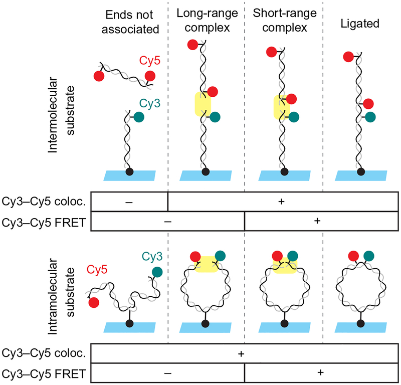
Fig. 2.
Schematic of intramolecular and intermolecular single-molecule DNA bridging assays. The intermolecular assay (upper panel) relies on a 100-bp DNA substrate labeled near one end with Cy3 (cyan circle) and tethered to the coverslip at the other end by a biotin–streptavidin attachment (black circle). This is incubated with egg extract containing a second 100-bp DNA labeled near both ends with Cy5 (red circles). Formation of the long-range synaptic complex (Graham et al., 2016) is detected in the inter-molecular assay based on the appearance of a discrete Cy5 spot that colocalizes with a Cy3-labeled DNA on the surface. Subsequent formation of the short-range synaptic complex is indicated by the appearance of FRET between Cy3 and Cy5. The intramolecular DNA substrate (lower panel) consists of a single, 2-kb DNA labeled near one end with Cy3 and near the other end with Cy5 and tethered to a streptavidin-coated coverslip via an internal biotin. Formation of the short-range synaptic complex (Graham et al., 2016) is indicated by appearance of FRET between Cy3 and Cy5.

