
Figure 2.
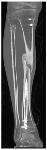
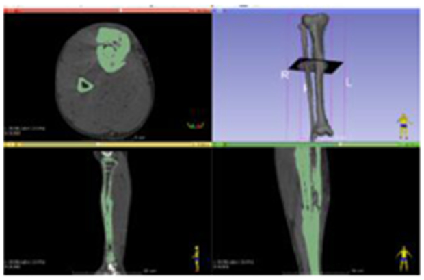
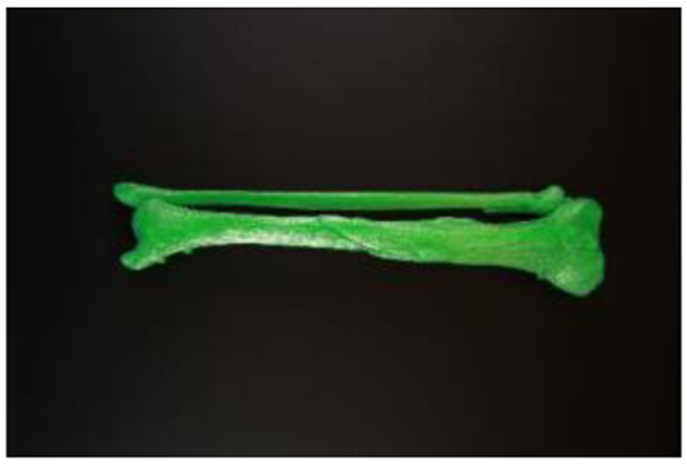
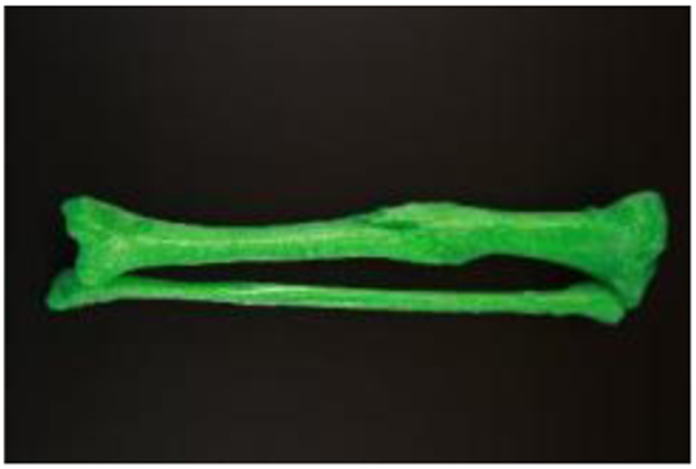
3D printed model of a tibial fracture nonunion. 42-year-old man 5 months following open reduction and internal fixation of a comminuted tibial fracture. a and b. Coronal reconstruction computed tomography (CT) image of the tibia and fibula with show a healed fibula fracture and a reduced and internally fixated, non-united tibial fracture. b. CT DICOM data used to create a STL file (3D Slicer version 4.6, www.slicer.org). Anterior (c) and posterior (d) photographs of a 3D printed anatomical model of the tibia and fibula show the orientation of the tibial fracture nonunion.