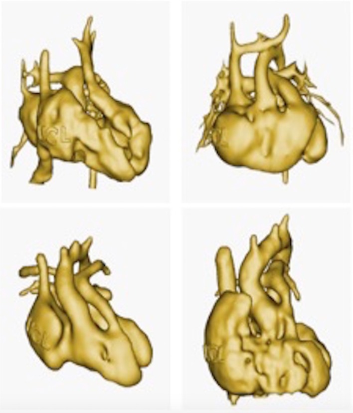
Figure 13.
The use of 3D model is particularly valuable for education in congenital cardiology and cardiac surgery. Patient anatomies are often unique and it is difficult to capture the wide range of anatomical variations that occur in the context of the same cardiac malformation. In this example, four patient specific virtual 3D models of double outlet right ventricle are imaged. (From 3D library at http://www.ucl.ac.uk/cardiac-engineering/research/library-of-3d-anatomies),

