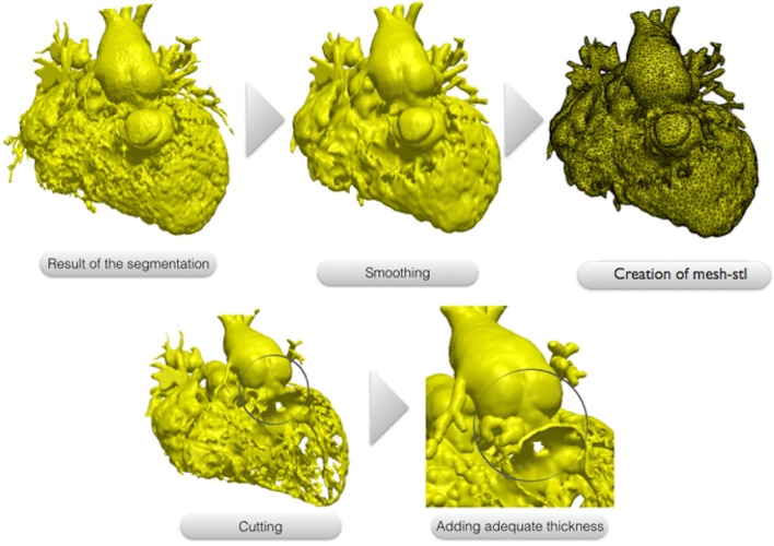
Figure 2.
The 3D images resulting from the segmentation process usually present a dyshomogenous surface due to the presence of multiple trabeculations, mostly inside the right ventricle. This file is modified though a smoothing tool using a commercial available software (ScanIP, Synopsis, US) and converted to a surface mesh file called “.stl”. The surface can be cut exposing the region of interest for the surgeon. In our case, the right and left ventricle free walls have been removed to allow the visualization of the multiple apical VSD. Finally a 1.2 mm thickness is added to the surface to make it suitable for printing.

