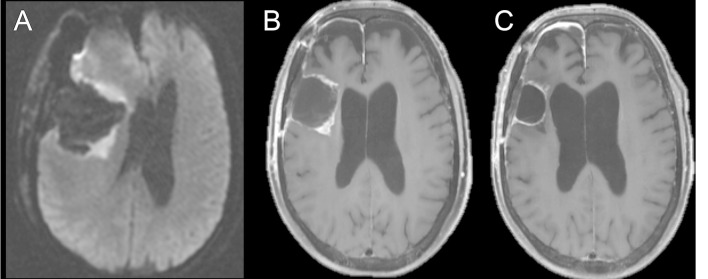
Figure 3.
Axial diffusion-weighted image of a patient with glioblastoma obtained immediately after surgical resection (A) shows restricted diffusion about the periphery of the surgical cavity. Axial T1 weighted post-contrast image (B) obtained 3 weeks after surgery for radiation planning demonstrates contrast enhancement in the areas of previous restricted diffusion, consistent with subacute enhancing infarction, which would otherwise be ambiguous without the previous DWI. Follow-up axial T1 weighted post-contrast image (C) demonstrates reduced contrast enhancement, consistent with evolution of post-surgical changes. DWI, diffusion-weighted imaging.

