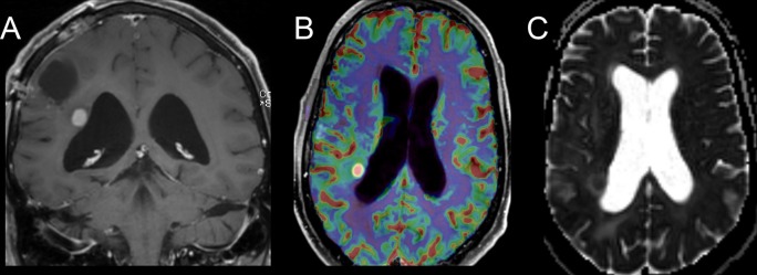
Figure 9.
Coronal T1 weighted post-contrast image (A) of a patient with glioblastoma demonstrates a focus of enhancement along the right lateral ventricle, deep to the resection cavity, which was new since the most recent comparison imaging. Perfusion MR image (B) demonstrates increased rCBV and the ADC map (C) shows restricted diffusion, both consistent with progressive, cellular, vascular tumor. ADC, apparent diffusion coefficient; rCBV, relative cerebral blood volume.

