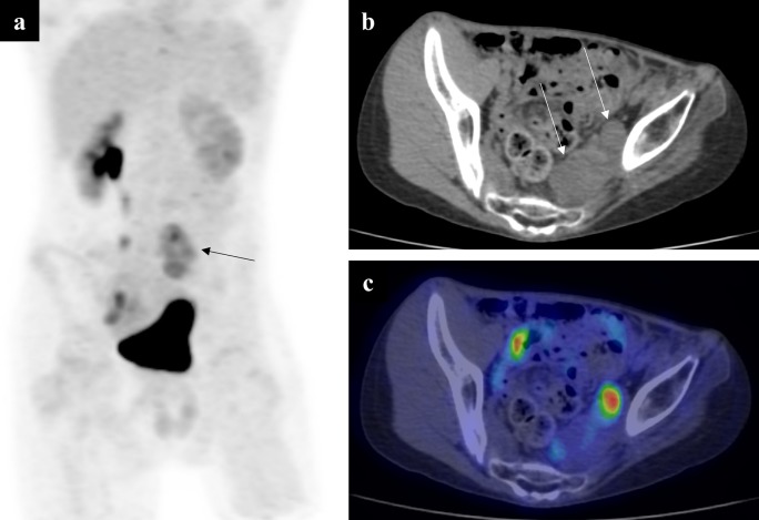
Figure 8.
A 13-year-old boy with a pelvic neurofibroma. The neurofibroma had previously shown malignant transformation and was treated. New symptoms of pain and left-sided lower limb neurology prompted an MR scan, which showed increase in the size of the residual neurofibroma (not shown). A PET MIP (a) showed a left-sided region of increased FDG uptake (arrow). Note the two discreet soft tissue masses on the low-dose CT component (b) at this level (arrows). On the fused axial PET-CT slice (c), the medial lesion has little-to-no FDG uptake, but the lateral mass shows high avidity. This imaging allowed a targeted biopsy to be performed, which showed sarcomatous transformation on histology. FDG, fluorodeoxyglucose; MIP, maximum intensity projection; PET, positron emission tomography.

