Figure 3.

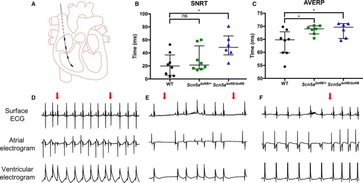
oElectrophysiological studies with programmed electrical stimulation in vivo. A, Schematic representation of electrical stimulation showing catheter with 4 electrodes inserted through the right jugular vein into the right atrium and right ventricle. Top electrode is used for atrial pace (“stimulating”) with the other 3 leads capture (“recording”) electrical activity in the heart. B, Duration of the sinoatrial node recovery time (SNRT) corrected for basic cycle length in Scn5a Δe6B/Δe6B and Scn5a Δe6B/+ mice compared with wild‐type (WT) littermates (P=0.013 and 0.465, respectively). C, Duration of the atrioventricular effective refractory period (AVERP) in Scn5a Δe6B/Δe6B and Scn5a Δe6B/+ mice compared with WT littermates (P=0.021 and 0.032, respectively). D, Representative intracardiac ECG tracing with premature atrial complex indicated by red arrow (4/5 Scn5a Δe6B/Δe6B, 0/8 WT). E, Representative intracardiac ECG tracing with sinus pauses indicated by red arrows (4/5 Scn5a Δe6B/Δe6B, 0/8 WT). F, Representative intracardiac ECG tracing with spontaneous reversion from bradycardia with the reversion indicated by the red arrow (3/5 Scn5a Δe6B/Δe6B, 0/8 WT). n=8 wild type, 8 Scn5a Δe6B/+, and 6 Scn5a Δe6B/Δe6B except for inducibility of arrhythmias where 1 died. *P<0.05.
