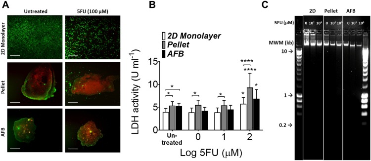
FIG. 7.
5FU induces cell death and apoptosis in Huh7 AFB generated discoids. (a) Live/dead staining of Huh7 cultured in the 2D monolayer, pellet, or AFB cultures 72 h following addition of 100 μM 5FU. Scale bar = 500 μm. (b) LDH (U ml−1) release in supernatants from Huh7 cultured in various culture conditions 72 h following addition of 5FU. n = 3. P values shown in the graph are for comparison to untreated cells (on top of bars) or to the cell in the 2D monolayer culture. *P < 0.05, **P < 0.005, ***P < 0.0005, ****P < 0.0001. Mean ± SEM. Two-way ANOVA followed by Fisher’s LSD test. (c) DNA fragmentation assay of Huh7 DNA following treatment with 5FU loaded in 1% agarose gel with different culture systems (representative image of 3 experiments). HyperLadder 1 kb molecular weight marker (MWM) was indicated.

