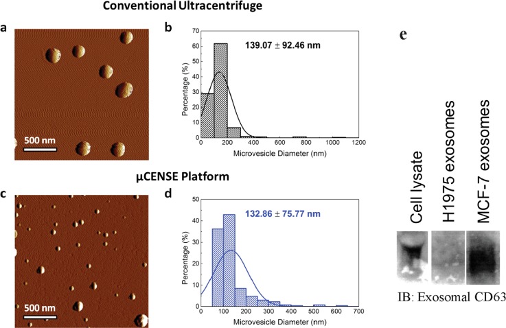
FIG. 5.
Comparison of extracted microvesicle separation using a commercial kit and the μCENSE platform. (a) Representative topological profile of microvesicles extracted from the conventional extraction kit using tapping mode atomic force microscopy. (b) Size distribution of the microvesicles (n = 287). (c) Representative topological profile of microvesicles extracted from the μCENSE platform using tapping mode atomic force microscopy. (d) Size distribution of the microvesicles (n = 270). The scale bar represents 500 nm. (e) Immunoblotting indicates the presence of exosomal CD63 markers in the microfluidic outlets for the MCF-7 cell line but absence in the H1975 cell line.

