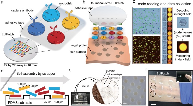
FIG. 1.
Schematic illustration of components and the usage of the ELIPatch. (a) ELIPatch has about 500 of encoded microdisks that have the shape code and corresponding antibody. Heterogeneous library of microdisks is placed in an array format with replicates on an adhesive tape. (b) Application of the ELIPatch to the human skin surface. (c) Automated data acquisition process. 10× images in bright field and dark field are used for decoding and fluorescence measurements. Scale bar, 500 μm. (d) Preparation of the ELIPatch by fluid-assisted self-assembly. Note that the size of the microwell fits to that of the microdisk. (e) Photographs of the ELIPatch in different scales. Microdisks in the 22 × 22 array are transferred to the 16 mm diameter tape by peeling off. Scale bar, 1 cm and 100 μm. (f) Example of application to Inner forearm. Black circles denote the array of ELIPatch. (inset) Size comparison with the ELIPatch and thumbnail. Scale bars, 1.5 cm.

