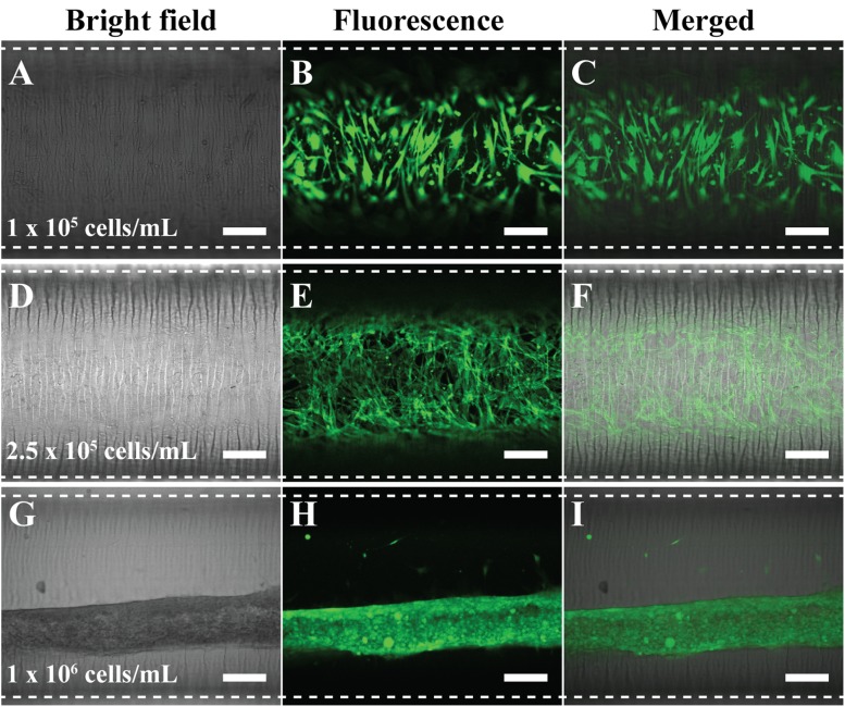
FIG. 2.
Optimized concentration of VSMC to fully cover inside the circular microfluidic channel. The used concentration was [(a)–(c)] 1 × 10⁵ cells/ml, [(d)–(f)] 2.5 × 10⁵ cells/ml, and [(g)–(i)] 1 × 10⁶ cells/ml. Filamentous actin of VSMCs was stained with a green fluorescence dye to be visualized. Dotted lines were the boundaries of the microfluidic channel. Scale bar: 200 μm.

