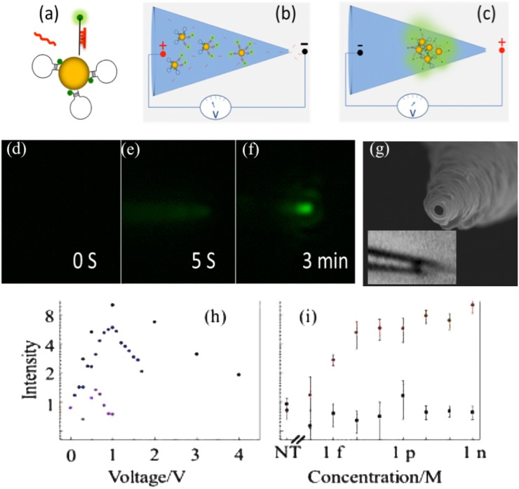
FIG. 6.
Hairpin Oligo Probe (HOP)-functionalized gold nanoparticles (AuNPs) nanopore-based detection. (a) Upon target hybridization, the fluorophore (green dot) is displaced beyond AuNP quenching distance. (b) Target miRNAs and HOP-AuNPs are driven into a conic nanocapillary by a negative voltage. (c) Voltage is reversed to aggregate AuNPs, promote hybridization, and plasmonically enhance fluorescence. (d)–(f) Microscopy sequence of HOP-AuNP packing and target miRNA hybridization in a silica conic nanocapillary; a negative voltage is applied at s and reversed at s; the plasmonically enhanced fluorescent signal is evident at min. (g) A nm nanocapillary tip SEM. Inset: light microscope image of a micron-sized nm-NP aggregate inside the silica nanocapillary. The inner nanocapillary diameter is about nm at the aggregate location; its distance from the tip ( ) corresponds to the ionic strength maximum in Fig. 9(a). (h) miRNA hybridization across a AuNP assembly in a conic glass nanocapillary: fluorescence intensity vs. voltage after target and non-target hybridization. (i) Fluorescent intensity for the target and for a -mismatch in the -nucleotide miRNA at different concentrations indicating little hybridization of the latter. Reproduced with permission from Egatz-Gomez et al., Biomicrofluidics 10(3), 032902 (2016). Copyright 2016 AIP Publishing LLC.

