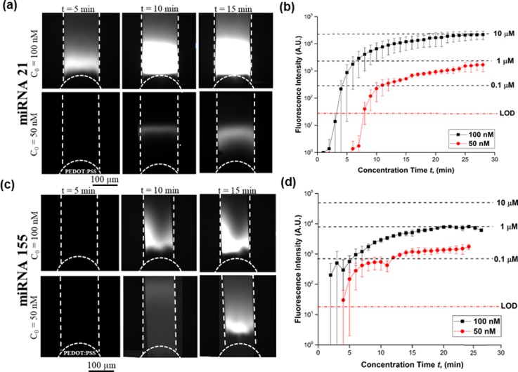
FIG. 2.
Characterization of miRNA concentrations 50 nM and 100 nM at 15 V. (a) Bright fluorescent plug inside the channel indicating miRNA 21 near the conductive polymer membrane—size continued to increase as a function of concentration time. (b) Fluorescence intensity for ND 1 filter and 1 s exposure time for miRNA 21. Within 30 min, the concentration of miRNA 21 increased by ∼90-fold for 100 nM and ∼20-fold for 50 nM. (c) Fluorescent plug inside the channel indicating miRNA 155 near the conductive polymer membrane. (d) Its concentration increased by ∼10-fold for 100 nM and ∼4-fold for 50 nM.

