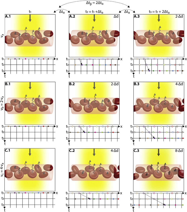
Fig. 3.
Illustration of how blood flow speed and interscan time affect OCTA signal. The transverse width of the OCT beam is shown in yellow. Black squiggly arrows indicate light backscattered from red blood cells. The rows correspond to different blood flow speeds (va, vb, and vc, increasing by factors 2x in this example) and columns correspond to three equally-separated time points of the repeated A-scans (t1, t2, and t3). The time between repeated A-scans is Δtα; the time between the first and third A-scans is Δtβ = 2 × Δtα. The left panels (A.1, B.2, and C.1) are all identical, representing the initial positions of the cells; while subsequent columns show the displaced blood cells (Δd, 2Δd, 4Δd). The graphs under each panel show how the positions of the cells (x) change with repeated A-scans (different colored dots showing different cells). The displacement of the blood cells depends on flow speed and interscan time (displacement = speed × interscan time). Measurements with twice the interscan time Δtβ = 2 × Δtα (i.e., first-to-third column) are equivalent to doubling the blood flow speed for an interscan time Δtα; thus, A.3 is identical to B.2, and B.3 is identical to C.2. When flow is fast vc and the interscan time is long Δtβ, a (purple) cell at the edge of the OCT beam and passes nearly to the other side (C.1 to C.3); this corresponds to the maximum distinguishable speed, or the saturation speed for that interscan time. Conversely, when flow is slow va and interscan time short Δtα, the cell translates very little during the interscan time (A.1 to A.2); this corresponds to the slowest detectable speed for that interscan time. By shortening the interscan time from Δtβ to Δtα (C.3 to C.2), the cell travels half the distance and so the saturation speed is halved and it is possible to distinguish differences in flow; conversely by lengthening the interscan time from Δtα to Δtβ (C.2 to C.3), the cell travels twice the distance and so the slowest detectable speed is halved and sensitivity is improved.

