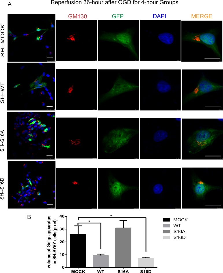
Fig 6. The morphological changes of the Golgi apparatus in SH-SY5Y cells after reperfusion for 36 hours following OGD for 4 hours in each group.
Ten cells in three images for each group were used for statistical analysis. (A) GM130, a stromal protein of the cis-Golgi apparatus, participates in maintaining Golgi morphology. Red represents GM130, green represents fluorescently labeled plasmids, and blue represents DAPI-labeled nuclei. After OGD/R, the Golgi apparatus fractured into many small fragments. The degree of Golgi apparatus fragmentation was reduced in the WT and S16D groups compared with the MOCK and S16A groups of human SH-SY5Y cells. The scale bar = 20 μm. (B) The GA volume in each group. * indicates P < 0.05.

