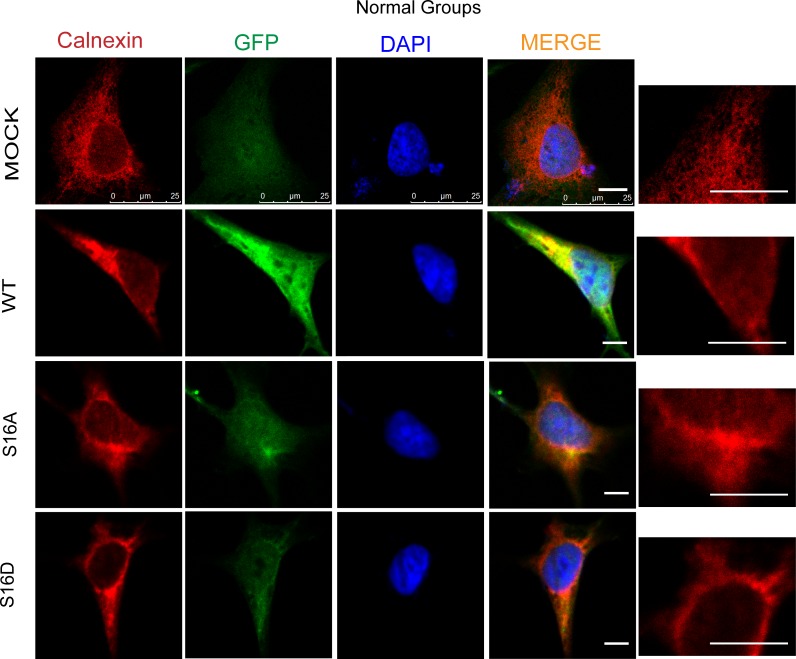
Fig 9. Endoplasmic reticulum morphology in SH-SY5Y cells transfected with pEGFP-N1, WT, S16A and S16D plasmids.
Red represents Calnexin, green represents fluorescently labeled plasmids, and blue represents DAPI-labeled nuclei; the far right is a magnified display of the endoplasmic reticulum. The scale bar = 10 μm.

