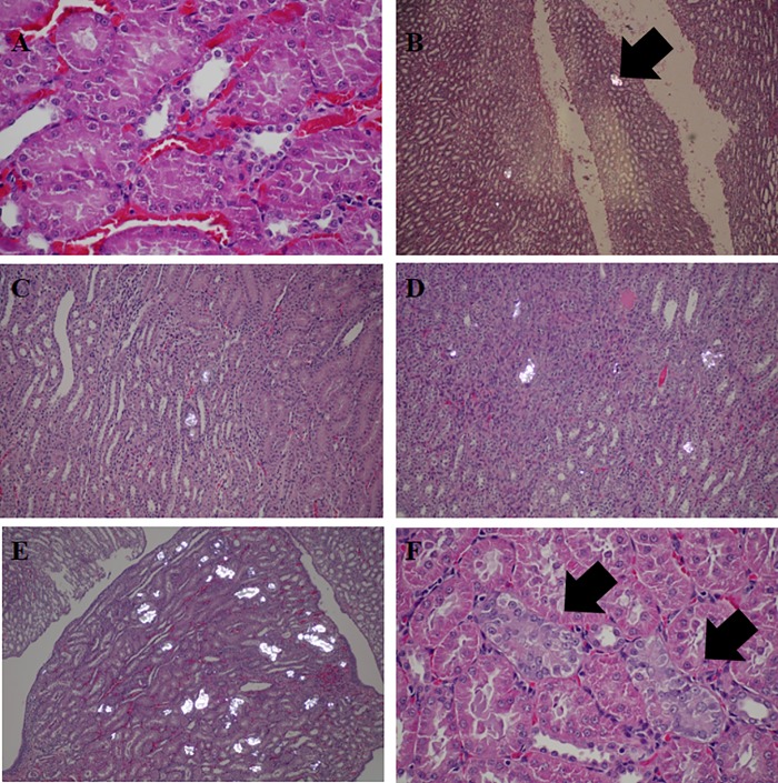
Fig 1. Urolithiasis grading in renal tissue with H&E staining visualized by light microscope (LM) and polarized microscope (PM).
Grade 0 contained calcium oxalate crystal deposit in all section (A, LM x40); Grade 1 contained less than 5 nephrons with calcium oxalate crystal deposition in distal tubular lumen (B, PM, x20); Grade 2 contained 5–19 nephrons with calcium oxalate deposition (C, PM x20); Grade 3 contained 20–50 nephrons with calcium oxalate deposition mostly in distal tubular regions, and occasionally in proximal tubular regions (D, PM x20); Grade 4 contained more than 50 nephrons with calcium deposition in both distal and proximal tubular regions (E, PM x 20); Tubular injury characterized by thin brush border, mildly enlarged nuclei, coarse chromatin and visible nucleoli was detected in the association with urolithiasis formation (E, LM x40).

