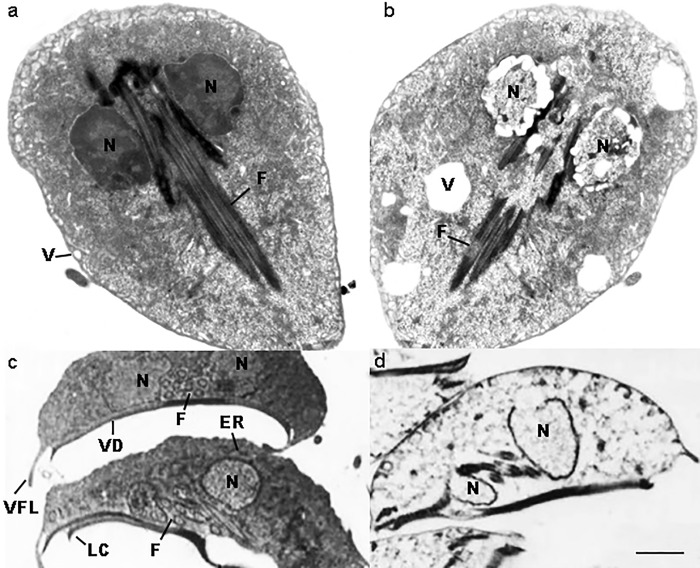
Fig 2. Ultrastructure of Giardia duodenalis by TEM.
Untreated G. duodenalis trophozoites showing normal structure and morphology (a). Trophozoite coronal section (c). A coronal view of a trophozoite demonstrates the nuclei (N), endoplasmic reticulum (ER), flagella (F), vacuoles (V), ventral disk (VD), lateral crest (LC) and ventrolateral flange (VLF). The same sections (b, d) of treated parasites show swelling trophozoites, with an increased size, and evident alterations of their cellular membrane and with a vacuolar degenerative pattern (x6,700). Note in the coronal section the severely damaged nuclei, nuclear membrane rupture, loss of the chromatin, flanges and ventral disk rupture (x6,700).

