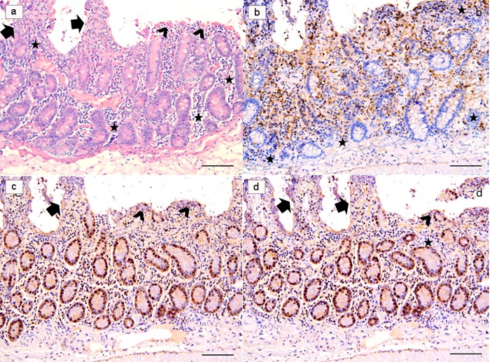
Fig 5. Ex-vivo intestinal tissue, from negative control (NC) group.
Section stained with H&E revealed a diffuse epithelial loss with inflammatory cells infiltration polarized under the destroyed intestinal epithelium. Note the reinforcement of inflammatory cells around intestinal glands (a). Villi are partially damaged (arrows), while in part they are totally flat or in any case strongly tuned (arrowheads) due to the effect of the strong colonization-adhesion of Giardia duodenalis trophozoites on the surface of the epithelium, which has become detached in many areas of the mucosa. Note the reinforcement of inflammatory cells around intestinal glands (stars). Ki67 nuclear staining evidenced an apparently higher number of positive cells because many inflammatory cells showed a strongly nuclear positivity (b). Note that TUNEL (c) and Caspase-3 (d) are over-expressed in these explanted intestinal samples. (H&E, and IHC with Mayer Haematoxilin nuclear counterstain, scale bar 400 μm).

