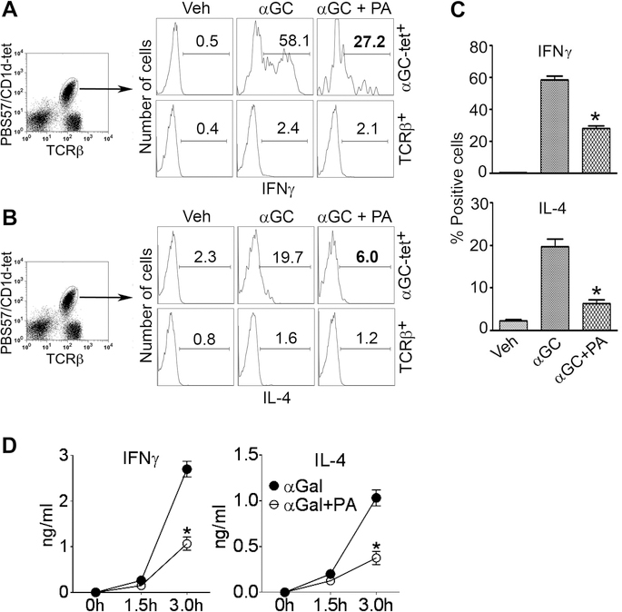
Figure 7. Effect of PA on cytokine production by iNKT cells.
B6 mice were injected with PA (20 μg i.p.) + αGalCer (2–4 μg i.p.) or αGalCer (αGC) alone, and animals were euthanized 3 h later. Liver MNCs were isolated and analyzed for intracellular cytokines. Representative histograms show IFNγ+ (A) and IL-4+ (B) cells on gated iNKT cells (PBS57/CD1d tetramer+ TCRβ+; upper panels) or conventional T cells (tetramer–TCRβ+; lower panels) from one of three independent experiments, each using 2 mice per group. Results are summarized in panel (C) (*p <0.004; n = 6 mice/group, pooled from three experiments; mean ± SE). (D) B6 mice were injected with αGalCer or αGalCer+PA, and animals bled at the indicated time points. Serum samples were assayed for cytokines by ELISA (*p =0.003; n = 6 mice/group, pooled from two separate experiments; mean ± SE).

